ABC Heart Fail Cardiomyop 2022; 2(4): 386-394
Critical Analysis and Applicability of Imaging Methods in Monitoring and Diagnosing Ventricular Dysfunction in Patients with Cancer
Claudio Tinoco Mesquita
![]() , Marcelo Dantas Tavares de Melo
, Marcelo Dantas Tavares de Melo
![]() , Ariane Binoti Pacheco Leal
, Ariane Binoti Pacheco Leal
![]() , André Luiz Cerqueira de Almeida
, André Luiz Cerqueira de Almeida
![]()
Abstract
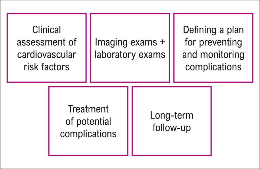
Cardio-oncology is a subspecialty of cardiology that has become necessary as a consequence of the favorable impact of cancer treatment, which increases patients’ survival rates, but makes them more prone to the cardiovascular side effects of cancer therapies in the short and long term. The presence of predisposing factors such as pre-existing cardiovascular disease, cardiovascular risk factors, genetic predisposition, previous antineoplastic therapies, and increased patient age is associated with a higher risk of cardiotoxicity in cancer treatment and may aggravate its complications. The use of imaging methods is fundamental in the management and detection of complications in cancer treatment. Echocardiography is considered the standard method for assessing left ventricular function and should be used in all patients. Magnetic resonance imaging is the best alternative for evaluation in patients with other associated conditions, especially advanced coronary disease, and in cases where it is difficult to obtain adequate echocardiographic images. Nuclear medicine offers options for patients for whom the use of echocardiography and magnetic resonance imaging is limited and for patients whose clinical and laboratory assessments conflict. Judicious use of imaging techniques leads to better patient outcomes during cancer treatment.
429