ABC Heart Fail Cardiomyop 2022; 2(3): 250-258
Myocarditis: Whole Heart Involvement Revealed by Cardiac Magnetic Resonance Mapping. A Case-control Study
Tiago Bartzen Pereira
![]() , Maurício Balk, Gabriela Bartzen Pereira, Victória Schmidt Ramos, Luciano Giordani, Paulo R. Schvartzman, Luís Beck-da-Silva
, Maurício Balk, Gabriela Bartzen Pereira, Victória Schmidt Ramos, Luciano Giordani, Paulo R. Schvartzman, Luís Beck-da-Silva
![]()
Abstract
Background:
Late gadolinium enhancement (LGE) on cardiac magnetic resonance (CMR) only demonstrates regional abnormalities in myocarditis and does not adequately assess diffuse myocardial involvement.
Objective:
To evaluate possible differences in T1 and T2 mapping between ventricular wall segments with and without LGE in patients with myocarditis, compared to control subjects.
Methods:
In a case-control design, 22 patients with CMR evidence of myocarditis and 18 controls with normal CMR were assessed. The study included: (1) T1 mapping (shortened modified Look-Locker Inversion recovery); (2) LGE; (3) T2 mapping (steady-state free precession); and (4) the T2 signal intensity of the myocardium divided by that of skeletal muscle (T2 ratio). T1 and T2 mapping of affected (LGE+) and unaffected (LGE−) ventricular segments of cases were compared, as were those of controls versus cases. The level of significance was set at a two-sided alpha level of 0.05.
Results:
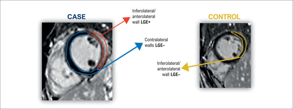
Comparing only patients with myocarditis, ventricular segments with evidence of late enhancement (LGE+) showed a mean T1 value significantly different from that of unaffected (LGE−) ventricular walls (1057 ± 30 versus 1028 ± 48; p = 0.0001). Comparing myocarditis versus controls, the mean T1 value of negative LGE segments in cases (myocarditis +) was significantly different from the mean of the corresponding walls in controls (1028 ± 48 versus 996 ± 10; p < 0.0001). The mean T2 maps of negative LGE walls in cases were not statistically different from those of controls (49 ± 4 versus 49 ± 1; p = 0.9229).
Conclusion:
This case-control study suggests that T1 mapping demonstrates significant involvement of the myocardium of patients with myocarditis, even in the absence of LGE. Specifically, T1 mapping could reveal diffuse myocardial involvement not evidenced by LGE imaging. T2 mapping was noncontributory.
Keywords: Contrast Media; Magnetic Resonance Imaging; Myocarditis
1,093